CARE-X Medical AI
Mild aortic dilation: 12% caught on first reads, 93% with a measuring VLM
Aortic dilation is rarely quantified on chest X-rays, and mild cases get missed: 5 of 43 on initial reads. A tool-augmented VLM caught 40. New CARE-X research explains why radiology AI needs rulers, not just eyes.
Emmanuel Fabrice Omgbwa Yasse AI-assisted
2026-08-16 · 5 min read
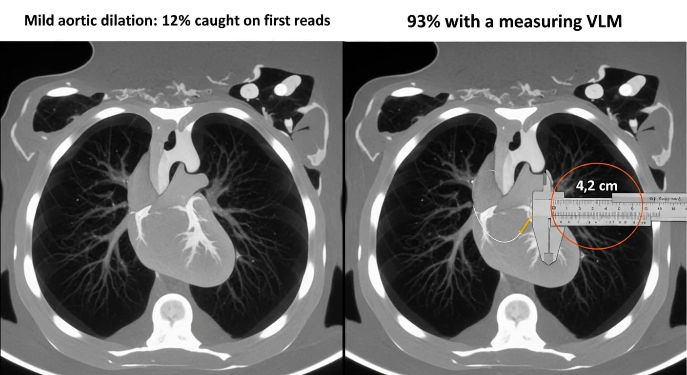
Five of 43. That is how many CT-confirmed cases of mild aortic dilation a set of initial chest X-ray reads caught in a study accepted at the EACTS conference in 2026. A measurement-driven AI approach found 40 of 43: 93% sensitivity against 12%. Aortic enlargement is usually not the primary indication for a chest X-ray, and it is not routinely quantified on one, so borderline cases stay invisible until a CT scan obtained for another indication happens to show them. The failure mode is not unique to chest X-rays: video language models also miss simple event bookkeeping. The research note ties delayed detection to adverse cardiovascular outcomes.
The model doing that measuring was not CARE-X. The experiment, a separate research effort described in the same note, paired Qwen3-VL-4B-Instruct with deterministic measurement tools. The VLM keeps visual access to the radiograph through a multi-turn loop, calling tools to identify anatomical landmarks, compute measurements, and evaluate diagnostic thresholds before answering. CARE-X is the companion piece: a unified chest X-ray vision-language model combining free-text report generation with calibrated structured predictions in a single forward pass.
When looking is not enough, add a ruler
Some radiological findings are defined by numbers, not patterns. Cardiomegaly means computing cardiac and thoracic widths and their ratio. Mediastinal widening, aortic knob enlargement, and dilated ascending or descending aorta all depend on measurements a model should calculate rather than approximate visually, while accounting for view, exposure, and patient rotation. With no task-specific training, the tool-augmented pipeline beat perception-only inference on every measurement-based condition evaluated, in line with research on when tool use helps multimodal models and when it hurts:
| Condition | Perception-only F1 | Tool-augmented F1 | Gain |
|---|---|---|---|
| Cardiomegaly | 74.56 | 96.00 | +21.4 |
| Mediastinal widening | 72.63 | 97.47 | +24.8 |
| Aortic knob enlargement | 60.31 | 99.76 | +39.5 |
| Ascending aorta enlargement | 39.33 | 100.00 | +60.7 |
| Descending aorta enlargement | 28.57 | 100.00 | +71.4 |
| Average | +43.6 |
The largest gains sit at the clinically dangerous end. Descending aorta enlargement went from 28.57 F1 with perception alone to a perfect 100 with tools; ascending aorta from 39.33 to 100. On a CT-confirmed outpatient cohort of 122 positive cases, the tool variant reached 94.26% recall, 10.65 points above the best perception-only baseline. In the EACTS study, the measurement-driven approach caught 40 of 43 mild aortic dilation cases against 5 of 43 on initial reads, surfacing 35 mild cases that had been missed. The design, deterministic tools bolted onto a neural model, echoes neuro-symbolic models beating raw neural ones in the TRACTA benchmark.
These figures measure recall only, and the authors say so. In triage, a missed diagnosis is usually the costlier failure, which is why the study focused on it, but a model that flags everything earns perfect recall and is useless in practice. An extended study with CT-confirmed negative cohorts is underway.
CARE-X: one model, two ways to answer
CARE-X is built on a SigLIP2-so400M vision encoder and a Phi-4-mini-instruct (3.8B) language model connected through a lightweight adapter, with task-specific auxiliary heads for classification and grounding co-trained alongside the language objective. Dual inference means one forward pass yields both an autoregressive text response and an auxiliary-head prediction carrying a confidence score. Because those scores are calibrated, thresholds can be moved: at 0.5 the model returns 0.943 sensitivity, at 0.6 it returns 0.927 positive predictive value. A clinician could shift between high-sensitivity screening and high-specificity confirmation from one pass, which purely generative architectures cannot provide.
Reinforcement learning with DAPO narrows the remaining gap between generation and structure, a bet shared by research on compiling standard operating procedures into executable code, which lifted agent performance by 16 points. On anatomical grounding in Chest ImaGenome, the DAPO-trained generative output hits 0.868 mAP, slightly ahead of the SFT detection head at 0.865, and the auxiliary grounding head lifts phrase grounding on PadChest by 24.6 points mAP and 14.1 points mIoU. CARE-X also ranked first on the ReXrank ReXVQA leaderboard as of August 2026, with 94% accuracy across 41,007 question-answer pairs, six points above the next-best publicly reported model, and posts the strongest performance on most metrics across MIMIC-CXR, IU-Xray, CheXpert-Plus, and ReXGradient.
Rare conditions on Indian clinical data
With Medha AI and Narayana Health as collaborators, the team validated CARE-X on 1,047 de-identified chest radiographs from Narayana Health, covering five rare, high-acuity conditions with prevalence between 2.6% and 5.2%. CARE-X posted the highest sensitivity in three of the five:
| Condition | CARE-X | CheXOne | MedGemma |
|---|---|---|---|
| Fracture | 0.62 | 0.41 | 0.05 |
| Mediastinal shift | 0.83 | 0.80 | 1.00 |
| Pneumoperitoneum | 0.89 | 0.67 | 0.00 |
| Pneumothorax | 0.83 | 0.85 | 0.52 |
| Tubes & lines abnormal placement | 0.66 | 0.03 | 0.18 |
Pneumoperitoneum came in at 0.89 sensitivity with 0.94 specificity. The widest gap is in abnormal tubes and lines placement: 0.66 against 0.03 for CheXOne and 0.18 for MedGemma. MedGemma's perfect 1.00 on mediastinal shift carries 0.53 specificity, the trade-off that makes raw sensitivity misleading on its own.
What the numbers do not say
None of this is ready for a hospital. CARE-X is a research model, not a Microsoft product offering and not a medical device. It has not been cleared or approved by any regulatory authority and is not intended for clinical diagnosis, screening, or patient care. The results are retrospective research findings that do not establish safety, effectiveness, or suitability for any clinical use. The measurement experiment is separate from CARE-X, and its strongest results cover only positive cases with CT-confirmed ground truth.
The road to a screening tool
The measurement direction already has a spotlight: the AI-based aortic dilatation screening application was selected as a finalist for showcase at the IHF Innovation Hub during the World Hospital Congress 2026. The CARE-X roadmap includes structured report generation, richer differential diagnosis support, tighter tool integration, and clinical context beyond the image, such as laboratory results and patient history.
For now, the durable finding is the division of labor. The VLM understands the image and locates the evidence; the tools do arithmetic on the landmarks. For threshold-dependent diagnoses, that separation is what turned a 12% detection rate into 93%. It is the same arrangement behind a circuit diagram model that pairs perception with symbolic solvers instead of guessing where the wires go.
- Source : Mild aortic dilation: 12% caught on first reads, 93% with a measuring VLM — 2026-08-11
Get the tech essentials in 3 minutes every morning
One email, every weekday, with what actually matters in AI and tech.